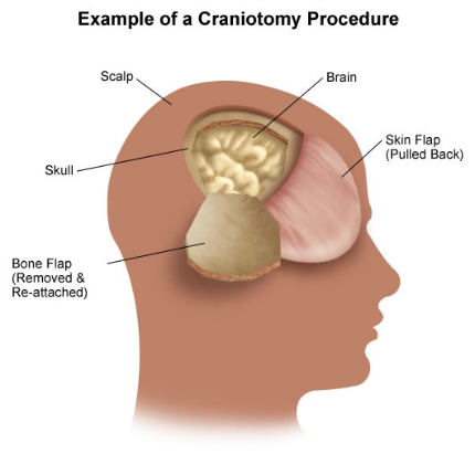
A craniotomy is the surgical removal of part of the bone from the skull to expose the brain. Specialized tools are used to remove the section of bone called the bone flap. The bone flap is temporarily removed, then replaced after the brain surgery has been done.
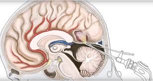
Some craniotomy procedures may use the guidance of computers and imaging (magnetic resonance imaging [MRI] or computerized tomography [CT] scans) to reach the precise location within the brain that is to be treated. This technique requires the use of a frame placed onto the skull or a frameless system using superficially placed markers or landmarks on the scalp. When either of these imaging procedures is used along with the craniotomy procedure, it is called stereotactic craniotomy.
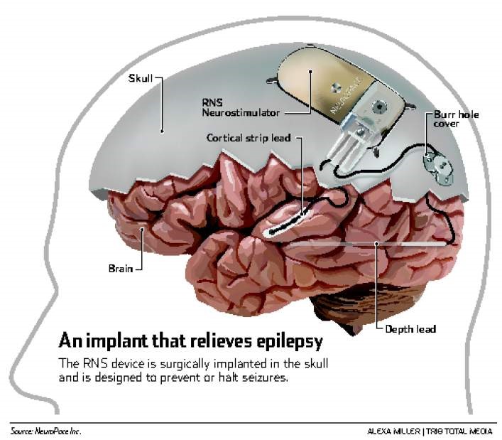
Burr holes are small holes that a neurosurgeon makes in the skull. Burr holes are used to help relieve pressure on the brain when fluid, such as blood, builds up and starts to compress brain tissue.
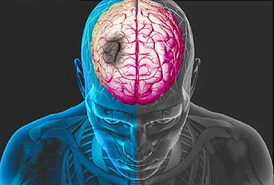
A layer of thin tissues called meninges surround and help protect the brain. These meninges contain blood vessels that carry blood to and from the brain. The dura is the outermost of these meninges. A head injury can cause one or more of these blood vessels to tear and bleed. A sudden tear might cause blood to build up very suddenly. With a small tear, the blood might build up more slowly. Blood might start to build up just below the dura mater. This causes something called a subdural hematoma. Tears in different blood vessels may cause blood to build up just above the dura layer, causing an epidural hematoma. A hematoma is when blood collects in an area and causes swelling.
This buildup of blood is dangerous. As the blood builds, it pushes up against the skull and has nowhere to go. If the blood starts to compress the brain, it can lead to symptoms or even death if not treated.
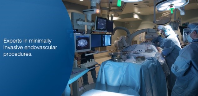

Numerous thrombogenic devices are available that can be delivered through microcatheters to permanently occlude cerebral vascular abnormalities. Catheter technology perfected in the coronary circulation for the management of atherosclerotic disease has been used to treat similar lesions of the extracranial and intracranial cerebral circulation.
Cerebral circulation angioplasty for vasospasm has become an accepted technique, and superselective drug delivery is growing in popularity. This chapter reviews the recent literature in vascular and endovascular neurosurgery in an attempt to familiarize the reader with recent advances in the management of cerebrovascular disease
WhatsApp us